
Redefining Wound Healing Using Near-Infrared Spectroscopy
The current standard of wound healing relies on what our eyes can see. What if what lies beneath the skin tells a different story?
Near-infrared (NIR) wavelengths penetrate ~2-3mm into the tissue to measure oxygenation saturation and hemoglobin levels. SnapshotNIR uses NIR light to monitor complete wound healing past visual inspection.
Andersen C, Reiter HJ, Marmolejo VL. Redefining Wound Healing Using Near-Infrared Spectroscopy. Adv Skin Wound Care. 2024 Feb 22. doi: 10.1097/ASW.0000000000000115. Epub ahead of print. PMID: 38408290.

Publication in Advances in Skin & Wound Care journal.
STUDY SUMMARY
Objective: This study aimed to fill an unmet clinical need for additional measurement tools to objectively assess wound healing progression. Historically, full wound healing requires 100% re-epithelialization on visual inspection.
This feasibility study aimed to add a clinical StO2 benchmark: a healed wound is when the tissue oxygenation of the wound matches the surrounding baseline tissue oxygenation.
Use objective data from Snapshot during each stage of patient care to improve outcomes.
Obtain SnapshotNIR assessment measures during initial patient evaluation and provide standard of care for the wound.
Monitor patient progress with Snapshot images at specified intervals.*
A wound closure image is to be taken on the day that 100% re-epithelialization occurs upon visual inspection.
Follow up Snapshot images should continue at specified intervals* until StO2 levels within the wound bed are similar to those of the surrounding, closed soft tissue envelope.
*Interval frequency is determined by the wound care provider for standard of care
Results: Monitoring 15 patients, with lower leg wounds and varying comorbidities, SnapshotNIR provided an objective measure that showed wound resolution occurred an average of 2 weeks, and up to 5 weeks, after 100% re-epithelialization on visual inspection.
Speak with confidence when advising patients on reloading the injured area and on return to activity to reduce risk of recurrence.
This study outlines how SnapshotNIR may help guide clinical decision-making for the optimal time for transition from protective wound dressings to protective shoe gear. Visualization of StO2 with point-of-care, noncontact, NIR imaging can assist clinicians in determining when deep dermal healing has occurred which can help guide patient education on gradual return to full activity to minimize wound recurrence.
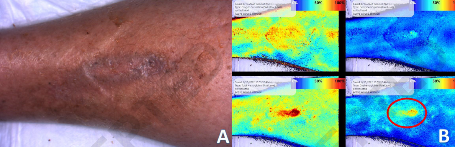
Study participant with a wound fully healed on visual inspection (A), compared to the SnapshotNIR image showing continued increased healing activity in the wound, particularly in the oxyhemoglobin frame (B).