
An algorithm that has positive impacts on health: Tissue oxygenation data for more skin tones
Health care disparities and skin assessment barriers in the under served and highly melanated patient population is a large concern in the industry.
How does melanin impact near-infrared spectroscopy?
Melanin is produced by melanocytes in the epidermis of our skin. When using near-infrared spectroscopy (NIRS), increased melanin content leads to a higher absorption of the NIR wavelengths reducing light reflectance. SnapshotNIR KD205, the latest device update, decreases the impact of melanin attenuating the NIR wavelengths, expanding the use of this technology to be used on more skin tones on any area of the body.

In the complexities of the American healthcare system, access to care and timely diagnosis continues to unequally burden marginalized populations. This is seen in conditions like peripheral arterial disease (PAD) which disproportionally impacts Black Americans and Hispanic American patients (1). This places these populations at higher risk for severe consequences such as minor and major amputations and premature death due to the complications from these diseases (1).
How does the snapshotnir technology work?
Kent Imaging has expanded SnapshotNIR’s technology to have the ability to capture repeatable and accurate objective data for tissue oxygen saturation (StO2) and hemoglobin, oxygenated and deoxygenated, in patients with varying levels of melanin content.
With these images from SnapshotNIR, clinicians can expedite referrals for vascular interventions and assist clinical judgement during healing chronic wounds. SnapshotNIR KD205’s proprietary algorithm can provide clinicians with the ability to assist in completing assessments and treatments across in patients almost all skin tones contributing to limb preservation and reducing healthcare costs.

Diagram of NIRS imaging through the layers of the skin and tissue. NIR wavelengths travel 2-3mm below the skin, through the dermis and the epidermis, where melanocytes are situated (2). Image adapted from Couch et al. 2015.
The near-infrared wavelengths of light used in SnapshotNIR KD205 are measured by the algorithm to determine levels of hemoglobin and calculate StO2. In individuals with darker skin, or more active melanocytes, the wavelengths are absorbed by melanin in the epidermis which can impact the amount of light reflected to the device (2), termed a light-scattering effect.
Light scattering can be impacted by:
skin melanin content
hair density
tissue collagen
fibrous skin components
SnapshotNIR Device Images - See Underneath the Skin
SnapshotNIR KD205’s proprietary algorithm addresses the light-scattering effect that high melanin content has on reflectance-based imaging devices.
The device calibrates and adjusts automatically to accurately measure StO2, oxyhemoglobin and deoxyhemoglobin in patients regardless of the level of melanin content in the skin to provide clinicians with repeatable, accurate data. Kent Imaging’s Snapshot KD205 update is a step towards advancing tissue assessment for physicians and patients. You can read more about SnapshotNIR KD205 in the full press release.
Below are clinical images using SnapshotNIR KD205 measuring StO2 and clinical, color images.
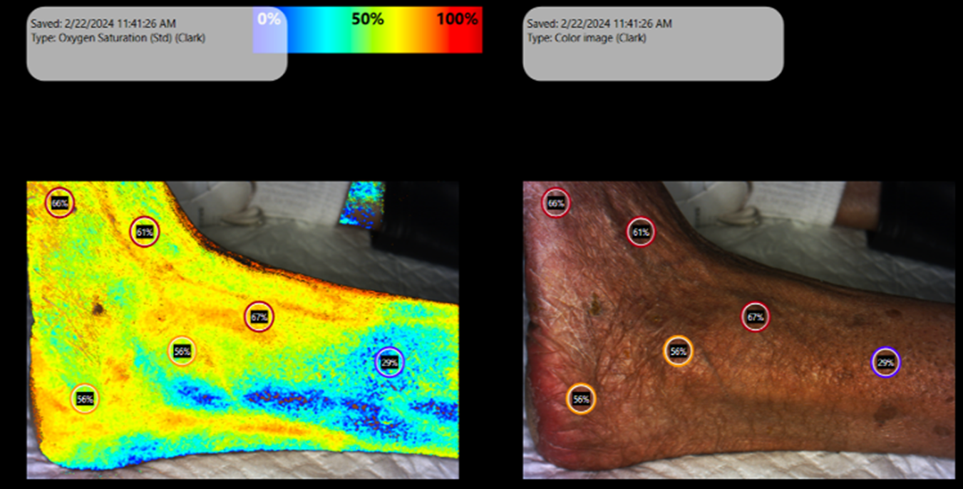
SnapshotNIR measuring tissue oxygen saturation on the lateral ankle. The markers on the ankle indicate the StO2 percentage.

Area measurement on the lateral toes of a patient using SnapshotNIR for monitoring wound size.

SnapshotNIR measuring tissue oxygen saturation for a breast reconstruction procedure. The markers indicate the StO2 percentage.

Linear wound measurement on the lateral plantar aspect of the patient’s foot.

Wound measurement using SnapshotNIR on a patient’s foot for accurate wound sizing.

SnapshotNIR measuring tissue oxygen saturation on an anterior leg. The markers indicate the StO2 percentage.
What do snapshotnir customers have to say?

Dr. Naz Wahab, MD
“SnapshotNIR has been extremely beneficial in our practice, particularly in individuals with darker complexions. I was able to diagnose anemia, I was able to diagnose people who had vascular issues, and people who perhaps also just had respiratory issues and were not oxygenating very well. Previously, it was very difficult to assess these conditions via tissue oxygenation in darker skinned patients.” - Dr. Naz Wahab MD, WCE Specialty
Watch the entire Q&A with Dr. Wahab below.
References:
Allison, Matthew A., et al. "Health disparities in peripheral artery disease: a scientific statement from the American Heart Association." Circulation 148.3 (2023): 286-296.
Couch, Luke, et al. "Effect of skin pigmentation on near infrared spectroscopy." American Journal of Analytical Chemistry 6.12 (2015): 911.